During vertebrate development, development of organs (organogenesis) is precisely regulated to elicit their functions. We are studying organogenesis and organ functions by using teleost species, such as zebrafish (Danio rerio) and medaka (Oryzias latipes).
Neural development-formation of cerebellum and cerebellar neural circuits
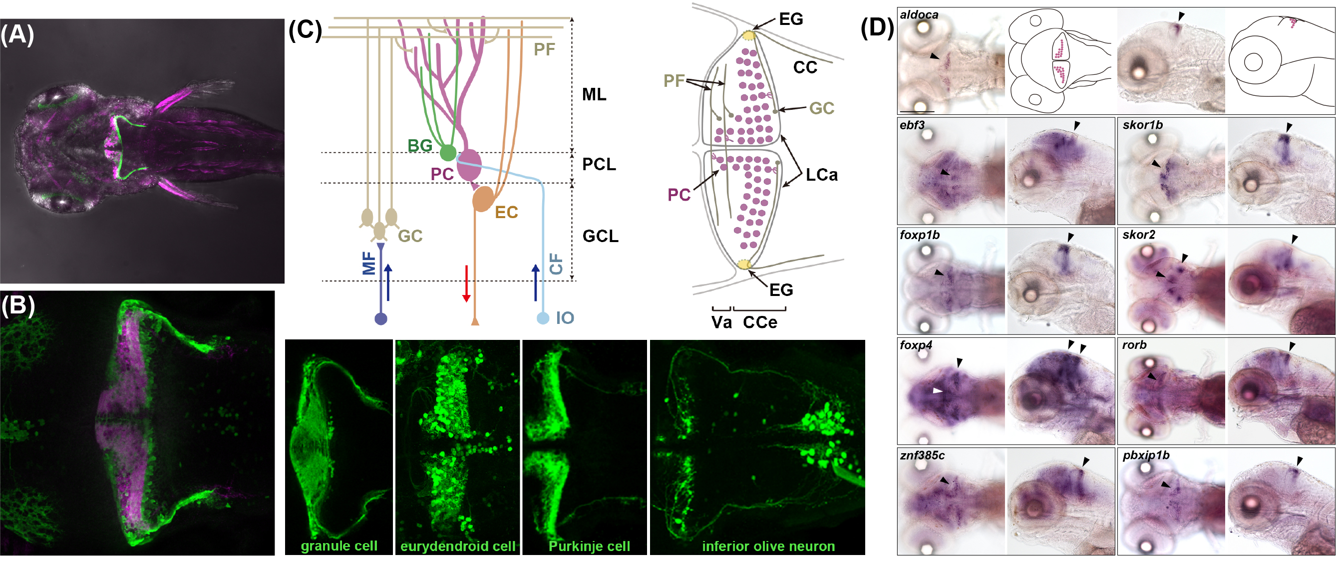
During neural development, the fate of neural tissues is determined along the rostro-caudal axis, depending on their location. In each neural tissue, neurons are generated from the neural stem cells and/or neuronal progenitors. Most of neurons concomitantly migrate and extending neurites, axons and dendrites that form neural circuits. As a model of neural development and functions, we currently focus on the cerebellum. The cerebellum is a neural tissue derived from the dorsal part of the most rostral hindbrain in the vertebrate central nervous system. The structure of the cerebellum and the cerebellar neural circuits is generally conserved among the vertebrates. There are two major cerebellar neurons, glutamatergic granule cells and GABAergic Purkinje cells, which are generated in distinct progenitor domains. Granule cells are derived from neuronal progenitors in the upper (cerebellar) rhombic lip that is located in the superficial domain of the cerebellum primordium. Purkinje cells are derived from neuronal progenitors in the ventricular zone that is located on the ventral side of the cerebellum primordium. They migrate, differentiate, and form neural circuits (Fig. 1). Granule cells and Purkinje cells receive afferent inputs, the mossy fibers (MFs) and climbing fibers (CFs), respectively, from outside the cerebellum. As the information of MFs is conveyed to Purkinje cells, the information of the MFs and CFs is integrated in Purkinje cells. Purkinje cells send outputs through the projection neurons (eurydendroid cells in teleost cerebellum) outside the cerebellum. This simple organization of the cerebellar neural circuits provides a good model to understand development of neural circuits in vertebrate CNS. We visualize the neural circuits by establishing transgenic zebrafish expressing exogenous protein, such as fluorescent proteins, and study developmental processes of the neural circuit formation. We further analyze molecular mechanisms that control differentiation of the cerebellar neurons and the input neurons (precerebellar neurons) by generating mutants with genome editing techniques. 【Topics】
Functions of neural circuits-classical fear conditioning
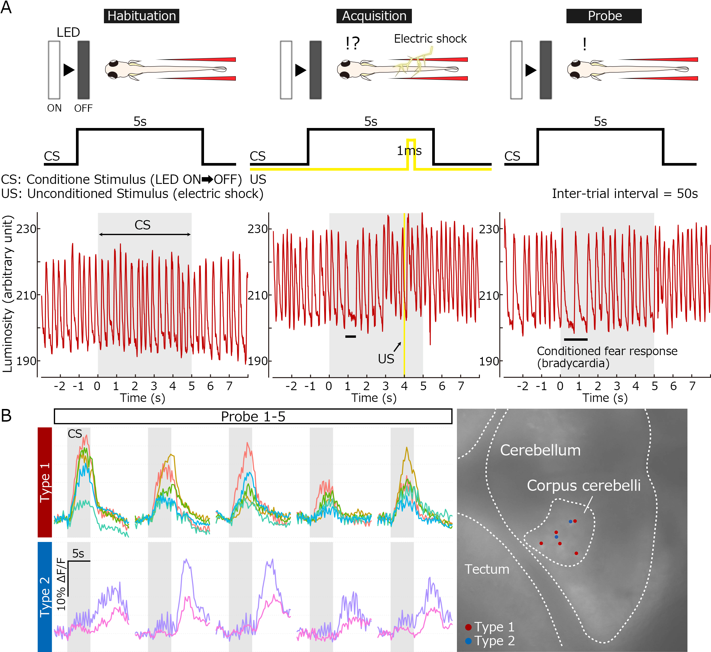
The cerebellum is known to function in some forms of skillful movements and motor learning. Recent studies reveal that the cerebellum is also implicated in higher cognitive and emotional functions. Furthermore, abnormalities in the cerebellar neural circuitry have been shown to be involved in pathogenesis of psychiatric disorders, such as autism. Therefore, elucidation of the functions of the cerebellar neural circuits provide some clue for understanding pathological conditions in related human diseases. We study roles of the cerebellar neural circuits in motor learning and fear conditioning by using zebrafish. We monitor the activity of the cerebellar neural circuits in the learning processes. We further examine their roles in the higher order functions by manipulating their activity using neurotoxin or optogenetic techniques. 【Topics】
Neural crest cells-differentiation of pigment cells
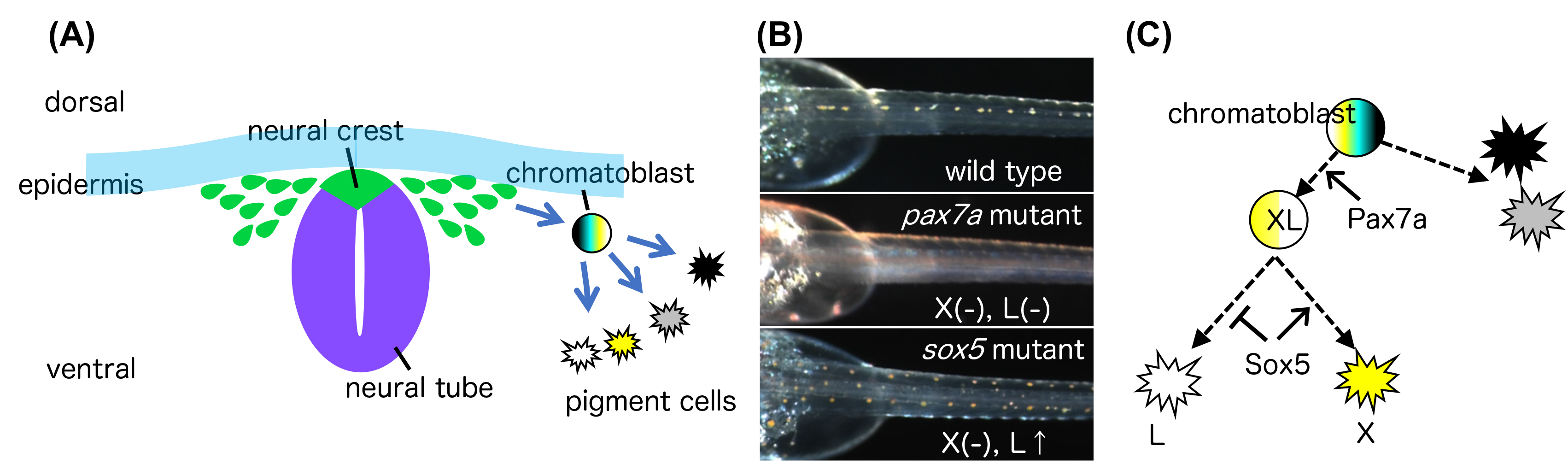
Pigment cells are derived from multipotent neural crest cells and their diversity in teleosts provides a good model for studying mechanisms controlling the specification of distinct cell types. Zebrafish have three types of pigment cells (melanophores, iridophores, and xanthophores), while medaka have four (three shared with zebrafish, plus leucophores). It remains elusive how different types of pigment cells are generated from the neural crest cells. In the late twentieth century, Hideo Tomita of Nagoya University isolated more than 80 naturally occurring medaka mutant strains called the Tomita collections. We have been studying Tomita-collection medaka mutants that display abnormalities in pigmentation. By establishing medaka and zebrafish mutants for key genes potentially involved in pigment cell differentiation, we study molecule mechanisms that control the pigment cell differentiation from the neural crest cells. By comparative studies with other teleosts, we try to understand evolution of the pigment cell development. 【Topics】
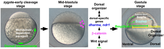
Axis formation-past research topics
The dorsal organizer plays a major role in the process of axis formation. In amphibian and teleost fish embryogenesis, the program for dorsal organizer formation begins soon after fertilization. In these species, the dorsal determinants are initially localized to the vegetal pole, and subsequently transported to the dorsal side of the embryo in a microtubule-dependent manner. The dorsal determinants activate the canonical Wnt pathway and thereby lead to the expression of the genes involved in the induction of the dorsal organizer. The dorsal organizer generates secreted signaling molecules that participate in the generation of the dorso-ventral axis. We have been studying the molecular mechanisms by which the dorsal organizer is established and regulates axis formation. We have also identified molecules that are involved in the formation of dorso-ventral, antero-posterior, and left-right axes. 【Topics】